
Carbon-13 NMR Spectroscopy: Interpreting HSC Chemistry Data
Learn how to count distinct carbon environments, use chemical shifts, recognise symmetry, and test proposed organic structures from carbon-13 NMR data.
A molecule can contain six carbon atoms but show only three peaks in a carbon-13 NMR spectrum. Another molecule with the same six carbons might show six peaks. The difference is not the number of carbons. It is the number of different carbon environments.
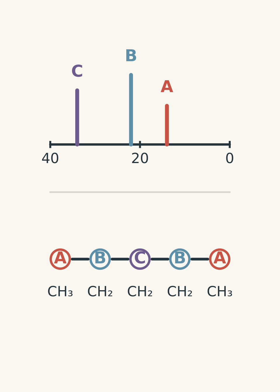
Before going further, predict this: would propane, \(\ce{CH3CH2CH3}\), give two carbon-13 NMR signals or three?
The two end carbons are chemically equivalent. Swapping the left and right ends does not create a different molecule, so those two \(\ce{CH3}\) carbons experience the same surroundings. The middle \(\ce{CH2}\) carbon is different. Propane therefore gives two carbon-13 NMR signals.
That simple idea – count distinct carbon environments, not carbon atoms – is the key to interpreting carbon-13 NMR in HSC Chemistry.
01What carbon-13 NMR is actually telling you
Most carbon atoms are carbon-12, which does not produce the NMR signal used here. A small proportion are carbon-13, written \(\ce{^{13}C}\), and these nuclei can interact with an applied magnetic field.
You do not need the detailed nuclear physics to interpret an HSC spectrum. The useful idea is this:
Carbon atoms in different chemical environments respond at different positions in the spectrum.
Each distinct carbon environment produces a signal at a particular chemical shift, measured in parts per million, or ppm.
So when you inspect a carbon-13 NMR spectrum, you mainly ask two questions:
- How many distinct carbon environments are present?
- What types of carbon could produce signals at those chemical shifts?
Those questions work together. Peak count tells you about the molecule’s symmetry and connectivity. Peak position tells you about the functional groups surrounding each carbon.
02One peak means one carbon environment, not one carbon atom
Imagine three students wearing the same school uniform. If two are standing in identical classrooms and one is standing in the chemistry lab beside a fume cupboard, they are not all experiencing the same surroundings.
Carbon atoms behave similarly. Two carbons count as the same environment only when the rest of the molecule surrounds them in an equivalent way.
Consider propan-2-ol:
\[ \ce{CH3CH(OH)CH3} \]
There are three carbon atoms, but the two \(\ce{CH3}\) groups are equivalent.
If you swapped them, nothing about the molecule would change. Therefore:
- the two methyl carbons give one signal,
- the central carbon gives another signal.
So propan-2-ol produces two carbon-13 NMR signals.
Now compare it with propan-1-ol:
\[ \ce{CH3CH2CH2OH} \]
The three carbons are all in different surroundings:
- one terminal carbon is part of \(\ce{CH3}\),
- one is the middle \(\ce{CH2}\),
- one is a \(\ce{CH2}\) directly bonded to oxygen.
It therefore gives three carbon-13 signals.
A useful symmetry test
When you are unsure whether two carbons are equivalent, imagine labelling one of them.
Would labelling the other carbon instead create a genuinely different arrangement?
If no, the carbons are usually equivalent.
If yes, they are different environments.
This is not a complete mathematical definition of molecular symmetry, but it is a very useful first test for the simple organic molecules encountered in HSC Chemistry.
03Chemical shift tells you what is near the carbon
Counting signals is only half the job. Their positions matter too.
An electron cloud partly shields a carbon nucleus from the external magnetic field. Electronegative atoms and certain bonding arrangements change the electron density around a carbon, changing its chemical shift.
The exact chemical shift depends on the whole molecular environment, so treat ranges as guides rather than rigid borders.
A practical HSC-level guide is:
| Approximate \(\ce{^{13}C}\) chemical shift | Carbon environment commonly found there |
|---|---|
| \(0\)-\(50\) ppm | Saturated carbon in alkyl groups |
| \(50\)-\(90\) ppm | Carbon bonded to an electronegative atom such as oxygen |
| \(100\)-\(150\) ppm | Carbon in many \(\ce{C=C}\) environments |
| roughly \(160\)-\(185\) ppm | Carboxylic acid and ester carbonyl carbons |
| roughly \(190\)-\(220\) ppm | Aldehyde and ketone carbonyl carbons |
These ranges overlap somewhat across real compounds and data tables. Use them as evidence alongside the molecular formula, functional-group tests, infrared spectroscopy, mass spectrometry, or other NMR data.
A signal near \(200\) ppm is particularly useful. A normal saturated alkyl carbon does not appear there. You should immediately suspect a carbonyl environment such as an aldehyde or ketone.
04Why nearby oxygen shifts a carbon signal
Suppose one carbon is in a simple alkyl environment, while another is directly bonded to oxygen.
Which do you expect to appear at the larger chemical shift?
The carbon attached to oxygen generally appears further downfield, at a larger ppm value.
Oxygen is highly electronegative. It draws electron density towards itself, so the attached carbon is less shielded from the applied magnetic field. That changes the resonance condition and gives a larger chemical shift.
The shorthand is useful:
electron-withdrawing surroundings tend to move a carbon signal downfield to higher ppm.
But do not turn that into a rule that chemical shift depends only on electronegativity. Double bonds, aromatic systems, and carbonyl groups also create important magnetic effects. The table works because it packages all of these effects into recognisable chemical-shift regions.
05Carbon-13 spectra are usually simpler than proton NMR spectra
In the carbon-13 spectra normally interpreted at this level, proton-carbon coupling is removed by proton decoupling.
That means each distinct carbon environment usually appears as a single peak rather than being split into the multiplets familiar from proton NMR.
So a typical carbon-13 NMR task is not about applying the \(n+1\) rule. It is mainly about:
- counting signals,
- recognising equivalent carbons,
- identifying chemical-shift regions,
- checking whether a proposed structure fits every signal.
If you are also working with proton NMR, the two techniques complement each other. Carbon-13 NMR tells you about distinct carbon environments, while proton NMR gives extra information about hydrogen environments, integrations, and neighbouring hydrogens. See Proton NMR Spectroscopy: How to Interpret HSC Chemistry Data for that side of the analysis.
06Worked example: How many carbon environments does butan-2-one have?
A compound has the structural formula \(\ce{CH3COCH2CH3}\). Predict the number of carbon-13 NMR signals and identify the likely region for each signal.
Step 1
There are four carbon atoms:
\[ \ce{CH3-CO-CH2-CH3} \]
Call them:
- carbon 1: \(\ce{CH3}\) beside the carbonyl,
- carbon 2: the carbonyl carbon,
- carbon 3: \(\ce{CH2}\) beside the carbonyl,
- carbon 4: the terminal \(\ce{CH3}\).
Step 2
The two \(\ce{CH3}\) carbons might initially look similar because they are both methyl groups. But their surroundings are different.
One is bonded directly to the carbonyl carbon:
\[ \ce{CH3-CO} \]
The other is bonded to \(\ce{CH2}\):
\[ \ce{CH2-CH3} \]
They are therefore different carbon environments.
All four carbons are distinct, so we expect four signals.
Step 3
The ketone carbonyl carbon should appear at about \(190\)-\(220\) ppm.
The remaining three carbons are saturated carbons and should appear much further upfield. The carbons close to the carbonyl are affected more strongly than the terminal methyl carbon.
A realistic spectrum might therefore contain signals around:
\[ \delta \approx 8,\ 30,\ 40,\ 205\text{ ppm} \]
where \(\delta\) is the chemical shift.
Step 4
Four peaks do not merely tell us that the molecule has four carbons. They tell us that the molecule contains four non-equivalent carbon environments.
The very high chemical shift near \(205\) ppm gives separate evidence for a ketone carbonyl.
07The most tempting mistake: counting every carbon
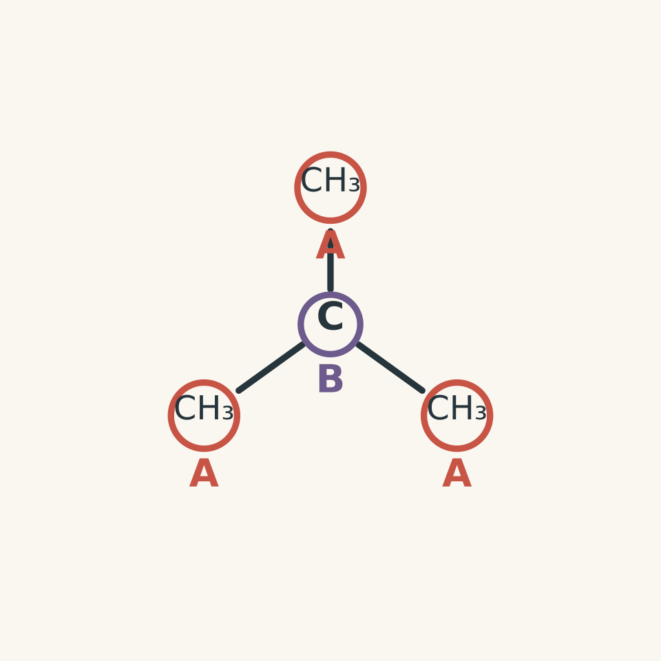
Consider 2-methylpropane:
\[ \ce{(CH3)3CH} \]
It contains four carbon atoms.
A student may therefore predict four signals. That feels reasonable if NMR is being treated as a carbon counter.
But the three methyl groups are equivalent. Each one has exactly the same relationship to the central carbon and to the rest of the molecule.
There are only:
- one methyl-carbon environment, containing three equivalent carbons,
- one central-carbon environment.
So the spectrum contains two signals, not four.
This is why symmetry matters so much. Carbon-13 NMR counts environments.
08Do peak heights tell you how many carbons are in each environment?
Not reliably in the usual carbon-13 spectra used for structural identification.
You might expect the signal representing three equivalent carbons to be three times the height of a signal representing one carbon. That would make carbon-13 spectra pleasantly convenient.
Unfortunately, ordinary proton-decoupled carbon-13 NMR peak intensities are affected by several experimental factors. They should not normally be treated as a direct count of carbons.
So this reasoning is unsafe:
“This peak is twice as tall, so it must represent two carbons.”
Instead, establish equivalence from the molecular structure and use the number and positions of signals as your main evidence.
09Worked example: Distinguishing two compounds with the same molecular formula
Two possible structures for a compound with molecular formula \(\ce{C4H10O}\) are butan-1-ol and 2-methylpropan-2-ol. A carbon-13 NMR spectrum contains only two signals, one near \(30\) ppm and one near \(70\) ppm. Which structure fits the spectrum?
Step 1
Its structure is:
\[ \ce{CH3CH2CH2CH2OH} \]
Each carbon has a different environment:
- terminal \(\ce{CH3}\),
- the next \(\ce{CH2}\),
- the next \(\ce{CH2}\),
- \(\ce{CH2OH}\).
So butan-1-ol should give four carbon signals.
The carbon directly bonded to oxygen should appear significantly further downfield than the ordinary alkyl carbons.
Step 2
Its condensed structure is:
\[ \ce{(CH3)3COH} \]
The three methyl carbons are equivalent because the molecule is symmetrical around the central carbon.
There are only two environments:
- the three equivalent methyl carbons,
- the central carbon bonded to oxygen.
So this structure should produce two carbon signals.
Step 3
A signal near \(30\) ppm is consistent with alkyl methyl carbons.
A signal near \(70\) ppm is consistent with a saturated carbon bonded to oxygen.
Therefore, the spectrum is consistent with 2-methylpropan-2-ol.
The important point is that both pieces of evidence agree:
\[ \text{two environments} + \text{a C bonded to O} \longrightarrow \ce{(CH3)3COH} \]
Peak count alone would narrow the options, but chemical shift makes the conclusion much stronger.
10A reliable method for interpreting carbon-13 NMR
When you are given a spectrum and asked to identify or assess a structure, use the evidence in this order:
- Count the signals. This gives the number of distinct carbon environments.
- Look for diagnostic regions. In particular, check for carbonyl carbons, carbons bonded to oxygen, and unsaturated carbons.
- Use the molecular formula if supplied. It limits the possible structures.
- Draw candidate structures.
- Mark equivalent carbons. Use symmetry rather than simply counting atoms.
- Predict one signal for each distinct environment.
- Compare every predicted environment with the observed chemical shifts.
- Reject a structure if even one important feature disagrees with the spectrum.
That final step matters. Structural analysis is not about finding one feature that agrees. A proposed molecule must explain the complete data set.
11Questions and solutions
Question 1
How many carbon-13 NMR signals would you expect from ethyl ethanoate, \(\ce{CH3COOCH2CH3}\)? Briefly identify the four carbon environments.
Solution 1
Ethyl ethanoate is expected to produce four carbon-13 NMR signals.
Its four carbons are all in different environments:
- the \(\ce{CH3}\) attached to the carbonyl carbon,
- the ester carbonyl carbon,
- the \(\ce{CH2}\) bonded to oxygen,
- the terminal \(\ce{CH3}\).
The carbonyl carbon should appear far downfield, typically in the ester carbonyl region around \(160\)-\(185\) ppm. The \(\ce{OCH2}\) carbon should also be shifted downfield compared with an ordinary alkyl carbon because it is directly bonded to oxygen.
A tempting mistake is to group the two \(\ce{CH3}\) carbons together. They are both methyl carbons, but they are not equivalent: one is next to a carbonyl group, while the other is next to \(\ce{CH2O}\).
Question 2
A compound has molecular formula \(\ce{C4H8O}\). Its carbon-13 NMR spectrum contains three signals at approximately \(22\), \(43\), and \(208\) ppm.
A student proposes butan-2-one, \(\ce{CH3COCH2CH3}\).
Is the proposed structure consistent with the spectrum? Explain fully.
Solution 2
No. Butan-2-one is not consistent with a spectrum containing only three carbon-13 signals.
The signal at approximately \(208\) ppm strongly supports the presence of a ketone carbonyl carbon, so that part of the proposal is sensible.
However, butan-2-one has the structure:
\[ \ce{CH3COCH2CH3} \]
Its four carbons occupy four different environments:
- \(\ce{CH3CO-}\),
- the carbonyl carbon,
- \(\ce{-COCH2-}\),
- terminal \(\ce{-CH3}\).
It should therefore produce four carbon-13 signals.
The trap is to notice that the \(208\) ppm signal fits a ketone and stop checking. Spectroscopic evidence has to fit the whole proposed structure. The incorrect number of carbon environments rules out butan-2-one despite the correct carbonyl region.
A structure such as 3-methylbutanal would not fit the molecular formula, so it cannot be substituted casually either. Both molecular formula and spectral evidence must be satisfied at the same time.
Question 3
Two structural isomers have molecular formula \(\ce{C5H10O}\).
Structure A is pentan-2-one:
\[ \ce{CH3COCH2CH2CH3} \]
Structure B is 3-methylbutan-2-one:
\[ \ce{CH3COCH(CH3)2} \]
A carbon-13 NMR spectrum shows four signals, including one at approximately \(205\) ppm.
Which structure is more consistent with the spectrum? Explain the role of symmetry.
Solution 3
Structure B, 3-methylbutan-2-one, is consistent with four signals; Structure A should give five.
Both compounds contain a ketone carbonyl, so the signal near \(205\) ppm is consistent with either structure. The decision therefore comes from the number of carbon environments.
For pentan-2-one,
\[ \ce{CH3COCH2CH2CH3} \]
all five carbons are in different environments. The two terminal methyl groups are not equivalent because one is attached directly to the carbonyl carbon while the other is at the opposite end of the chain.
So Structure A should give five signals.
For 3-methylbutan-2-one,
\[ \ce{CH3COCH(CH3)2} \]
the two methyl groups attached to the \(\ce{CH}\) carbon are equivalent. Interchanging them does not change the molecule.
Its distinct environments are therefore:
- the methyl carbon attached to the carbonyl carbon,
- the carbonyl carbon,
- the \(\ce{CH}\) carbon,
- the two equivalent methyl carbons attached to that \(\ce{CH}\).
That gives four signals.
The tempting route is to count five carbon atoms in each molecule and expect five peaks from both. The symmetry in Structure B removes one distinct environment, so only four signals appear.
Question 4
A compound has molecular formula \(\ce{C6H12O}\). Its carbon-13 NMR spectrum contains only three signals:
- \(24\) ppm,
- \(52\) ppm,
- \(211\) ppm.
A student argues:
“The \(211\) ppm peak means there is a ketone. Six carbons and only three signals probably means every signal represents two carbons.”
Assess this reasoning and propose a structure consistent with the data.
Solution 4
The ketone conclusion is reasonable, but the claim that each signal must represent exactly two carbons is incorrect. A consistent structure is 2,2-dimethylbutan-3-one, equivalently written as \(\ce{CH3COC(CH3)3}\), whose preferred name is 3,3-dimethylbutan-2-one.
Consider the structure:
\[ \ce{CH3COC(CH3)3} \]
It contains six carbons but only three distinct environments:
- the carbonyl carbon,
- the methyl carbon on the \(\ce{CH3CO-}\) side,
- the three equivalent methyl carbons in the \(\ce{C(CH3)3}\) group.
So the environments contain \(1\), \(1\), and \(3\) carbons respectively, not two carbons each.
The signal near \(211\) ppm is consistent with a ketone carbonyl.
The remaining signals near \(24\) and \(52\) ppm are consistent with saturated carbon environments influenced to different degrees by the nearby carbonyl group.
The student’s mistake is subtle. If six total carbons produce three signals, an average of two carbons per signal can be calculated, but an average does not determine the actual grouping. Carbon-13 NMR signal count tells us only that there are three distinct environments. One environment may contain three equivalent carbons while another contains only one.
The deeper constraint is symmetry: equivalent carbons must be related by the molecular structure, not assigned to groups merely to make the arithmetic come out neatly.
Question 5
An unknown compound has molecular formula \(\ce{C4H8O2}\). Its carbon-13 NMR spectrum contains only two signals: one near \(21\) ppm and one near \(170\) ppm.
A student proposes methyl propanoate, \(\ce{CH3CH2COOCH3}\), because an ester carbonyl should occur near \(170\) ppm.
Another student proposes ethanoic anhydride:
\[ \ce{CH3CO-O-COCH3} \]
Using only the molecular formula and carbon-13 NMR information given, determine which proposal is consistent with the evidence. Explain why the more obvious functional-group match is not enough.
Solution 5
Ethanoic anhydride is consistent with the two-signal spectrum; methyl propanoate is not.
The peak near \(170\) ppm indicates a carbonyl environment typical of carboxylic-acid derivatives such as esters or anhydrides. So methyl propanoate initially seems plausible.
But its structure must also reproduce the number of carbon environments.
For methyl propanoate,
\[ \ce{CH3CH2COOCH3} \]
there are four distinct carbon environments:
- terminal \(\ce{CH3}\),
- \(\ce{CH2}\),
- carbonyl carbon,
- methoxy carbon, \(\ce{OCH3}\).
It should therefore produce four carbon-13 signals, including a carbon attached directly to oxygen that would normally occur well above the simple alkyl region.
That does not match the observed two signals.
Now consider ethanoic anhydride:
\[ \ce{CH3CO-O-COCH3} \]
The molecule is symmetrical.
The two carbonyl carbons are equivalent, so together they produce one signal near \(170\) ppm.
The two methyl carbons are also equivalent, so together they produce one signal near \(21\) ppm.
Therefore:
\[ 4\text{ carbon atoms} \rightarrow 2\text{ distinct carbon environments} \rightarrow 2\text{ signals} \]
which fits the data.
The tempting mistake is to identify the \(170\) ppm signal as “an ester peak” and immediately choose an ester. Chemical shifts usually identify a type of environment, not an entire molecular structure. Closely related functional groups can occupy overlapping regions.
The decisive evidence here is the interaction between chemical shift and symmetry. The carbonyl signal narrows the functional-group possibilities, while the unusually small number of signals demands a highly symmetrical arrangement.
12What carbon-13 NMR lets you do next
Carbon-13 NMR becomes much more powerful when you stop treating it as a stand-alone identification trick.
A spectrum can tell you how many distinct carbon environments a candidate structure must contain and what broad types of carbon are present. You can then combine that with molecular formula, infrared data, mass spectra, and proton NMR to eliminate structures one constraint at a time.
The useful habit is simple: before naming a compound, make your proposed structure predict the spectrum. Count its environments, identify its expected chemical-shift regions, and check that every important observation fits.