
How to Choose a Radioisotope for Medical and Industrial Use
Learn how to select a radioisotope for medical imaging or industrial radiography by comparing radiation type, penetration, detectability, and half-life. Includes PET explained, worked examples, and HSC-style practice questions with solutions.
A doctor wants to detect a tiny radioactive tracer inside a patient’s body. An engineer wants to find a crack hidden inside a thick steel weld. Both need radiation to reach a detector, but the right radioisotope for one job might be useless for the other.
Before reading on, predict what matters more when choosing a source: its half-life, or the type of radiation it produces? The radiation and detector must be compatible first. Half-life then determines whether enough activity remains for the task, from preparation through to the end of the measurement.
Requested apparatus schematic is unsupported
| Medical imaging | Requires an internal tracer, escaping photons, and an external detector. |
|---|---|
| Industrial weld inspection | Requires an external radioactive source, a cracked steel weld, and a detector measuring transmitted radiation. |
01The decision rule
Use these criteria in order whenever you need to select a radioisotope for medical imaging or industrial use.
- Match the radiation to the detector. A gamma camera detects gamma photons, while a PET scanner detects coincident pairs of photons produced by positron annihilation.
- Check penetration. The radiation must pass through enough of the body or material to reach the detector, without losing so much contrast that the result becomes difficult to interpret.
- Match the half-life to the schedule. The isotope must remain sufficiently active during transport, preparation, and measurement.
- Check the activity and practical constraints. Consider the initial activity, expected signal, radiation dose, shielding, source lifetime, and how the isotope behaves in its intended setting.
If an isotope fails one of the first two criteria, a longer half-life or a higher initial activity won’t necessarily make it suitable.
01Start with the radiation and the detector
An isotope’s radiation type determines how it interacts with matter. That interaction determines whether the radiation can escape the object being studied and whether the detector can register it.
Which types of radiation are useful?
Radiation penetration: requested drawing unsupported
| Alpha particles | Very short range; readily stopped by tissue or thin barriers. |
|---|---|
| Beta particles | Travel farther than alpha particles but can be absorbed by sufficient tissue or metal. |
| Gamma photons | Generally more penetrating; attenuation depends on photon energy and material thickness. |
| Radiation | Penetration and detection | Implication for selection |
|---|---|---|
| Alpha particles | Highly ionising, with a very short range in matter | Unsuitable for external imaging through a patient’s body or thick metal |
| Beta-minus particles | Electrons that can be absorbed by tissue or metal before reaching an external detector | Usually a poor choice for external imaging of a deep target |
| Gamma rays | Penetrating electromagnetic radiation that can escape the object and reach a detector | Useful for SPECT and industrial gamma radiography |
| Positrons (beta-plus particles) | Travel a short distance through matter before annihilating with electrons | Useful for PET because the annihilation produces detectable photons |
Notice that positrons have a special role. A PET scanner does not need the positron itself to escape from the body. Instead, it detects the photons produced when the positron annihilates with an electron.
PET: how a positron becomes a detectable signal
PET stands for positron emission tomography. It uses a positron-emitting radioisotope attached to a tracer that follows a particular biological process.
Consider fluorine-18 (F-18), a commonly used PET radionuclide. Its physical half-life is about 110 minutes.
PET: from positron emission to coincidence detection
- Fluorine-18 decay
An F-18 nucleus emits a positron (β⁺).
- Positron slows
The positron travels a short distance through tissue, losing kinetic energy.
- Electron–positron annihilation
At the annihilation point, a positron meets an electron; their mass energy produces two photons.
- Coincidence detection
Photon 1 and photon 2 each carry approximately 511 keV and travel in nearly opposite directions towards detectors on opposite sides of the detector ring.
The process works as follows:
- The F-18 nucleus emits a positron.
- The positron loses kinetic energy as it travels through the surrounding tissue.
- The positron encounters an electron and the two particles annihilate.
- Their rest-mass energy is converted into two photons, each with an energy of approximately 511 keV, travelling in nearly opposite directions.
The PET scanner contains detectors arranged around the patient. When two detectors register suitable photons in coincidence, the scanner identifies a line along which the annihilation probably occurred. Many such events are combined to reconstruct an image of the tracer’s distribution.
The important distinction is that F-18 produces positrons, but PET detects the annihilation photons. The photons must still escape the body and reach the detector, and some will be absorbed or scattered along the way.
Industrial gamma radiography: radiation must cross the material
Now move the source outside the object.
In industrial gamma radiography, a radioactive source sends gamma photons through a metal component towards a detector on the opposite side. The detector records the transmitted radiation, producing an image of differences within the material.
Industrial gamma radiography: attenuation reveals defects
| Shielded gamma source | Emits penetrating gamma photons towards the weld. |
|---|---|
| Sound steel | More material along the beam path generally means greater attenuation and fewer transmitted photons. |
| Void or crack | Less material along the path can mean less attenuation and more transmitted photons; crack orientation affects visibility. |
| Digital detector | Records transmitted radiation; differences in signal may reveal defects. |
Why does a crack matter? A void can reduce the amount of material along a radiation path, allowing more photons to reach the detector than along a path through intact metal. The resulting difference may reveal the defect.
However, a penetrating source does not automatically reveal every crack. A narrow planar crack may produce very little contrast if the beam crosses its small opening rather than travelling along its plane. Material thickness, beam direction, detector resolution, and exposure conditions all affect whether a defect is visible.
Two examples of industrial gamma-emitting isotopes are iridium-192 (Ir-192) and cobalt-60 (Co-60). Both can be used for industrial radiography, but they are not interchangeable in every situation.
02Penetration, photon energy, and detectability
Knowing that an isotope emits gamma rays is only the starting point. You must also consider the energy of those photons and the material they must cross.
A low-energy photon may be absorbed before it reaches the detector. A more energetic photon is generally more penetrating, which can make it useful for imaging thicker objects.
But higher energy is not automatically better. If photons pass through a material with too little difference between sound and defective regions, the image may have insufficient contrast. Higher-energy sources can also increase shielding requirements.
The goal is to obtain a detectable signal with enough contrast to answer the question being investigated.
Medical imaging versus industrial use
| Criterion | Medical imaging | Industrial gamma radiography |
|---|---|---|
| Source position | The radioisotope is introduced into the patient as part of a tracer | The radioactive source remains outside the object |
| Required signal | Radiation escaping the body, or annihilation photons for PET | Gamma photons transmitted through the material |
| Penetration | Enough to escape tissue and reach the detector | Enough to pass through the relevant metal thickness |
| Half-life | Long enough for preparation and scanning, but not unnecessarily persistent | Long enough for transport, repeated inspections, and the intended service life |
| Additional constraints | Tracer distribution, patient dose, and biological clearance | Image contrast, source shielding, access control, and long-term source management |
The same physical principle applies in both cases: the detector must receive a useful signal. The difference is where the source sits, what the detector measures, and what the radiation needs to cross.
Don’t confuse activity with photon energy
Activity measures how many nuclear decays occur per second. Its unit is the becquerel (Bq), where \(1\text{ Bq}=1\) decay per second. One megabecquerel (MBq) is \(10^6\) decays per second.
A source with a higher activity generally produces more decays per second. However, activity does not tell you how energetic each photon is, whether the photons can escape the material, or whether a particular detector can measure them.
The number of useful detected photons also depends on the probability of emitting the relevant radiation, attenuation, detector efficiency, and measurement geometry.
This is why an isotope with a higher activity can still be the worse choice.
03Half-life and exposure duration
Once the radiation type and detector are compatible, consider how quickly the source loses activity.
The half-life, \(T_{1/2}\), is the time taken for the activity of a radioactive sample to fall to half its initial value through radioactive decay.
The activity after an elapsed time is calculated using
\[ A(t)=A_0\left(\frac12\right)^{t/T_{1/2}} \]
where:
- \(A(t)\) is the activity remaining after time \(t\), measured in Bq or MBq.
- \(A_0\) is the initial activity, measured in the same units.
- \(t\) is the elapsed time.
- \(T_{1/2}\) is the half-life.
Keep the time units consistent. If the half-life is given in hours, express the elapsed time in hours before substituting.
The exponent \(t/T_{1/2}\) tells you how many half-lives have passed.
- After one half-life, \(50\%\) of the initial activity remains.
- After two half-lives, \(25\%\) remains.
- After three half-lives, \(12.5\%\) remains.
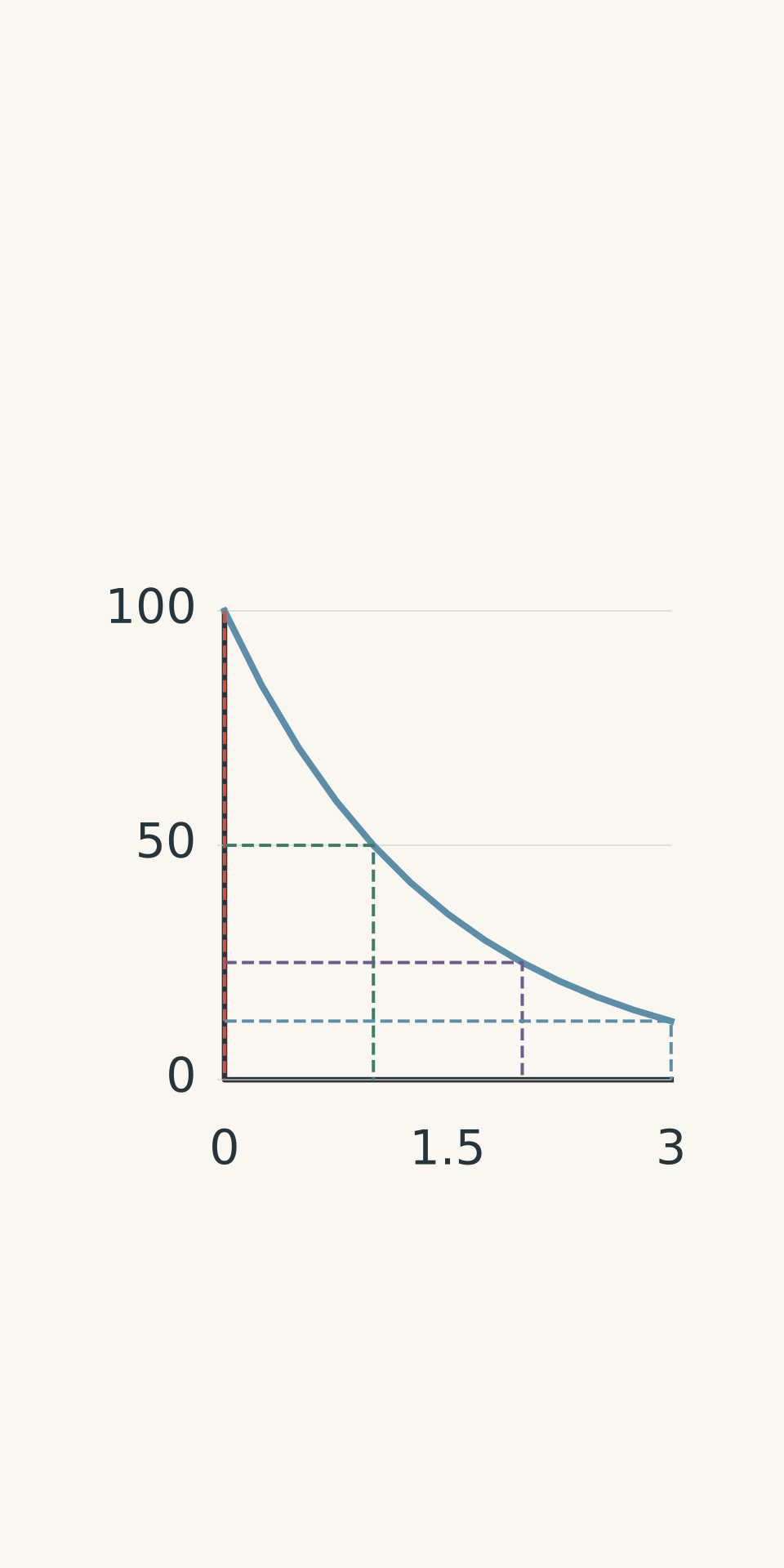
- Physical decay
- Initial activity: 100%
- 1 half-life: 50%
- 2 half-lives: 25%
- 3 half-lives: 12.5%
Why the task duration matters
Compare the half-life with the entire time between preparing the source and finishing the measurement.
For medical imaging, this includes transport, preparation, administration, and scanning. A short-lived isotope may be useful because its activity decreases relatively quickly, but it may also lose too much activity before the scan finishes.
For industrial radiography, an exposure might last minutes, while the source remains in service for months or years. A source’s activity may barely change during one exposure even though it decreases substantially over its service life.
Here are some useful approximate reference values:
- Technetium-99m (Tc-99m): half-life of about 6 hours; emits gamma photons, including a prominent photon near 140 keV. Commonly used for SPECT imaging.
- Fluorine-18 (F-18): half-life of about 110 minutes; emits positrons for PET imaging.
- Iridium-192 (Ir-192): half-life of about 74 days; emits gamma rays for industrial radiography.
- Cobalt-60 (Co-60): half-life of about 5.27 years; emits high-energy gamma rays, including photons near 1.17 MeV and 1.33 MeV.
These are starting points, not automatic selections. The appropriate isotope still depends on the detector, target, geometry, and radiation-safety requirements.
Worked example: choosing a tracer for an external gamma camera
A hospital needs to image a deep organ using a conventional gamma camera. Candidate X emits beta-minus particles and has a half-life of 8.0 hours. Candidate Y emits gamma photons near 140 keV and has a half-life of 6.0 hours. Both have an initial activity of 300 MBq, and the scan begins 2.0 hours after preparation.
Which candidate is the better match, and what activity remains at the start of the scan?
Step 1
Candidate Y is the better match. A gamma camera detects gamma photons that escape the body. Beta-minus particles from candidate X are much more likely to deposit their energy in tissue before reaching an external detector.
Step 2
Use the radioactive decay equation:
\[ \begin{aligned} A_X(2.0\text{ h}) &=300\text{ MBq} \left(\frac12\right)^{2.0/8.0}\\ &=300\text{ MBq}\times0.8409\\ &\approx252\text{ MBq} \end{aligned} \]
Step 3
\[ \begin{aligned} A_Y(2.0\text{ h}) &=300\text{ MBq} \left(\frac12\right)^{2.0/6.0}\\ &=300\text{ MBq}\times0.7937\\ &\approx238\text{ MBq} \end{aligned} \]
Step 4
Candidate X retains more activity, approximately 252 MBq compared with 238 MBq for candidate Y. Yet candidate Y is the better choice for this detector because its gamma photons can provide the required external signal.
This is the key lesson: having more activity is not the same as having a more useful signal. In a real medical application, the tracer’s chemical behaviour, biological distribution, and radiation dose would also need to be assessed.
Worked example: choosing a source for thick steel
An industrial team must radiograph a thick steel casting. For this example, assume that Ir-192 does not provide sufficient transmission through the casting, while Co-60’s higher-energy gamma photons can. Both sources currently have an activity of 1.0 TBq.
Compare their remaining activities after 148 days, assuming physical decay only, and explain the selection.
Step 1
Co-60 is the more plausible choice under the stated conditions. Its higher-energy gamma photons are better suited to the required penetration through the thick steel.
Step 2
Ir-192 has a half-life of approximately 74 days. Therefore, 148 days represents two half-lives.
\[ \begin{aligned} A_{\mathrm{Ir}}(148\text{ d}) &=1.0\text{ TBq} \left(\frac12\right)^{148/74}\\ &=1.0\text{ TBq}\times\frac14\\ &=0.25\text{ TBq} \end{aligned} \]
Step 3
First, convert its half-life into days:
\[ T_{1/2}=5.27\times365 \approx1924\text{ days} \]
Now substitute into the decay equation:
\[ \begin{aligned} A_{\mathrm{Co}}(148\text{ d}) &=1.0\text{ TBq} \left(\frac12\right)^{148/1924}\\ &\approx1.0\text{ TBq}\times0.948\\ &\approx0.95\text{ TBq} \end{aligned} \]
Step 4
After 148 days, Ir-192 has about 0.25 TBq remaining, while Co-60 has about 0.95 TBq. Co-60 retains substantially more activity and meets the stipulated penetration requirement.
However, Co-60 is not automatically the best source for every industrial inspection. Its high-energy photons increase shielding demands and may provide less useful contrast for some material and defect combinations. The detector, geometry, and required image quality must still be checked.
The assumed transmission requirement in this example is specific to the question. There is no single thickness boundary that determines the best isotope for every inspection.
04Common mistakes
Mistake: Choosing the isotope with the longest half-life.
A long half-life helps a source retain activity over an extended period. It does not make the radiation more detectable or more suitable for the target. A long-lived source also remains radioactive for longer and requires appropriate long-term controls.
Mistake: Choosing the shortest half-life because it must be safest.
A short half-life can reduce how long radioactive material persists, but it may also cause the signal to become too weak before the measurement finishes. Radiation dose depends on more than half-life, including initial activity, radiation energy, emissions, and where the tracer accumulates.
Mistake: Assuming gamma rays always produce a useful image.
Gamma photons can be absorbed or scattered, and the detector must be able to register the relevant signal. PET requires coincident annihilation photons; detecting individual gamma photons is not the same measurement.
Mistake: Assuming higher activity means greater penetration.
Activity measures decays per second. Photon energy and interactions with the material determine penetration. Increasing activity does not make each photon more energetic.
Mistake: Checking activity only at the beginning of a scan or exposure.
If the half-life is comparable with the task duration, activity may change substantially during the measurement. Calculate the remaining activity at the relevant time, and consider how the changing signal affects the measurement.
06Questions and solutions
Question 1
A Tc-99m tracer has an activity of 200 MBq at 08:00. Its half-life is 6.0 hours.
Calculate its activity at 20:00, assuming physical decay only. Explain why Tc-99m is useful for SPECT imaging.
Solution 1
The activity is 50 MBq at 20:00, and Tc-99m is useful for SPECT because its gamma photons can escape the body and reach an external detector.
The elapsed time is 12 hours, so the number of half-lives is
\[ n=\frac{12\text{ h}}{6.0\text{ h}}=2 \]
Using the decay equation,
\[ \begin{aligned} A(12\text{ h}) &=200\text{ MBq} \left(\frac12\right)^2\\ &=200\text{ MBq}\times0.25\\ &=50\text{ MBq} \end{aligned} \]
The activity halves twice: from 200 MBq to 100 MBq, then to 50 MBq.
For SPECT, the gamma camera detects photons emitted by the tracer. The tracer’s distribution provides information about the biological process being investigated. The calculated activity describes physical decay only; it does not include biological clearance from the patient.
Question 2
An engineer uses gamma radiography to inspect a steel weld. A highly penetrating Co-60 source is placed on one side of the weld, and a detector is placed on the other. The resulting image shows no obvious crack.
A student concludes that the weld must be sound because Co-60 gamma rays can penetrate thick steel.
Explain why this conclusion is unjustified. Describe two changes to the inspection geometry or method that could improve the chance of detecting a crack.
Solution 2
The absence of a visible crack does not prove that the weld is free of defects. Penetration allows radiation to reach the detector, but a defect must also produce a detectable difference in the transmitted signal.
A narrow planar crack may be difficult to see if the beam crosses its small opening rather than travelling along its plane. The difference between the radiation transmitted through the cracked region and the surrounding material may then be too small to distinguish clearly.
Two improvements are:
- Change the beam angle and take another projection. A different orientation may cause the radiation path to sample more of the crack, increasing the difference in transmitted intensity.
- Use a suitable alternative inspection method or improve the imaging conditions. The team could use an appropriately selected detector and exposure procedure, or a complementary non-destructive testing technique suited to the suspected defect.
The tempting assumption is that a highly penetrating source must reveal every internal flaw. In reality, penetration and defect detectability are different requirements. The result depends on contrast, geometry, resolution, and the inspection method as well as the source.
Question 3
An F-18 sample has an activity of 320 MBq at 07:00. Its half-life is 110 minutes.
A PET scan begins at 08:50 and finishes at 09:45.
Assuming physical decay only, calculate the activity at the beginning and end of the scan. Explain how F-18 produces the signal measured by the PET scanner.
Solution 3
The activity is 160 MBq at the beginning of the scan and approximately 113 MBq at the end. F-18 is suitable for PET because its positrons produce annihilation photons that the scanner detects in coincidence.
From 07:00 to 08:50, 110 minutes have elapsed. This is one half-life.
Therefore,
\[ \begin{aligned} A(110\text{ min}) &=320\text{ MBq} \left(\frac12\right)^1\\ &=160\text{ MBq} \end{aligned} \]
From 07:00 to 09:45, 165 minutes have elapsed. This represents
\[ n=\frac{165}{110}=1.5 \]
half-lives.
The activity at the end is
\[ \begin{aligned} A(165\text{ min}) &=320\text{ MBq} \left(\frac12\right)^{1.5}\\ &\approx320\text{ MBq}\times0.3536\\ &\approx113\text{ MBq} \end{aligned} \]
The activity falls noticeably during the scan because the half-life is of a similar order to the measurement duration.
In PET, the emitted positron slows down and annihilates with an electron, producing two photons of approximately 511 keV each. The scanner detects suitable photon pairs in coincidence and uses many such events to reconstruct the tracer’s distribution.
The calculation does not establish whether a real scan would have sufficient counts. Detector sensitivity, tracer uptake, photon attenuation, and biological clearance also affect the usable signal.
Question 4
Two hypothetical industrial gamma sources have the same initial activity of 800 MBq and emit photons with the same energy and emission probability.
- Source A has a half-life of 20 minutes.
- Source B has a half-life of 10 days.
A single industrial exposure lasts 40 minutes.
Calculate the activity of each source at the end of the exposure. Identify which source maintains a more stable output, and evaluate the claim that the source with the shorter half-life must give workers a lower radiation dose.
Solution 4
Source A finishes at 200 MBq, while source B finishes at approximately 798 MBq. Source B maintains a much more stable output, but half-life alone does not determine worker dose.
For source A, the exposure lasts two half-lives:
\[ \begin{aligned} A_A(40\text{ min}) &=800\text{ MBq} \left(\frac12\right)^{40/20}\\ &=800\text{ MBq}\times0.25\\ &=200\text{ MBq} \end{aligned} \]
For source B, convert the half-life into minutes:
\[ 10\text{ days}\times24\times60 =14400\text{ min} \]
Then,
\[ \begin{aligned} A_B(40\text{ min}) &=800\text{ MBq} \left(\frac12\right)^{40/14400}\\ &\approx798.5\text{ MBq} \end{aligned} \]
Source B’s activity changes very little during the 40-minute exposure. Source A’s activity falls to one-quarter of its initial value.
However, the apprentice’s claim is not justified. Source A still emits radiation throughout the exposure, particularly during its early stages. The total radiation emitted over the whole exposure depends on how activity changes over time, not merely on the final activity.
Under the stated conditions, source B produces more total emissions during the exposure because it remains active for longer. The actual dose received by a worker also depends on shielding, distance, exposure time, occupancy, and the procedure used.
A short half-life may reduce the activity remaining later, but it does not automatically make a source safer during a particular task.
Question 5
A remote imaging clinic has access to a PET scanner that accepts only suitable coincident pairs of photons near 511 keV.
Two candidate radioisotopes are available:
- Isotope P: emits gamma photons directly, with a photon energy of 140 keV and a half-life of 6.0 hours.
- Isotope Q: emits positrons, with a half-life of 110 minutes.
Both have an activity of 800 MBq when dispatched. Transport and setup take 3.0 hours, followed by a 2.0-hour scan.
For this hypothetical protocol, the activity must be at least 150 MBq at the end of the scan. Assume only physical decay, ignore biological clearance, and treat the stated activity threshold as the protocol’s requirement rather than a clinical recommendation.
Determine whether either isotope meets both the detector and activity requirements on this schedule. If neither does, calculate how much earlier the scan must finish for isotope Q to meet the activity requirement, assuming the scan still lasts 2.0 hours. Explain why simply choosing the isotope with more activity remaining is not a valid solution.
Solution 5
Neither isotope meets both requirements on the stated schedule. Isotope P retains more activity but cannot provide the required PET signal, while isotope Q produces the correct signal but falls below the activity threshold. The total elapsed time must be reduced by approximately 34 minutes for isotope Q to meet the stipulated requirement.
The key is to check detector compatibility and remaining activity separately.
Step 1: Calculate the total elapsed time.
Transport and setup take 3.0 hours, and the scan takes another 2.0 hours.
\[ t=5.0\text{ h}=300\text{ min} \]
Both isotopes must be assessed at this time because the requirement applies at the end of the scan.
Step 2: Calculate the activity remaining for isotope P.
Its half-life is 6.0 hours, so
\[ \begin{aligned} A_P(5.0\text{ h}) &=800\text{ MBq} \left(\frac12\right)^{5/6}\\ &\approx449\text{ MBq} \end{aligned} \]
Isotope P remains above 150 MBq. However, its directly emitted 140 keV gamma photons are not the coincident 511 keV annihilation photon pairs required by this PET scanner.
Therefore, P fails the detector requirement despite retaining plenty of activity.
Step 3: Calculate the activity remaining for isotope Q.
The half-life is 110 minutes, giving
\[ \begin{aligned} A_Q(300\text{ min}) &=800\text{ MBq} \left(\frac12\right)^{300/110}\\ &\approx121\text{ MBq} \end{aligned} \]
Isotope Q is the correct type for PET because positron annihilation produces the required photons. However, its activity falls below the stipulated 150 MBq threshold by the end of the scan.
Thus, Q has the right radiation mechanism but fails the timing requirement as scheduled.
Step 4: Determine the latest allowable finishing time.
For Q to finish with at least 150 MBq, set the remaining activity equal to the threshold:
\[ 150=800\left(\frac12\right)^{t/110} \]
Rearranging gives
\[ \begin{aligned} t&=110\log_2\left(\frac{800}{150}\right)\\ t&\approx266\text{ min} \end{aligned} \]
The total elapsed time must therefore be no more than about 266 minutes, rather than the current 300 minutes.
The required reduction is
\[ 300-266\approx34\text{ min} \]
Since the scan itself must still last 120 minutes, transport and setup must take no more than about 146 minutes, approximately 34 minutes less than the current 180 minutes.
Alternatively, the clinic could consider a compatible positron-emitting isotope with a longer half-life, provided it also meets the tracer and imaging requirements. Increasing the initial activity could satisfy the mathematical threshold, but that is not automatically an appropriate clinical decision because patient dose must be considered.
The tempting choice is P because it retains substantially more activity. The decisive constraint is that P cannot supply the signal this PET scanner is designed to measure. Q has the correct signal but needs a workable schedule.
The selection must satisfy both constraints at once: the radiation must be detectable, and enough activity must remain for the measurement.
07What to check next
When solving a new radioisotope selection problem, start by identifying the detector and the path the radiation must travel. Then check penetration and contrast, calculate the remaining activity at the relevant time, and consider the safety and practical constraints of the application.
The next useful step is to distinguish physical half-life from biological clearance. In medical imaging, the radionuclide can decay while the tracer is also being removed from the body. Understanding both processes helps explain why the activity remaining in a sample is not necessarily the same as the signal available from a patient.