
Mass Spectrometry of Organic Compounds: HSC Chemistry Guide
Learn how to interpret mass spectra by using molecular ions and fragment peaks to identify structural information about organic compounds.
A forensic chemist finds a tiny sample of an unknown organic liquid. The sample is too small to burn, and there is not enough material for a full set of tests. One question comes first: what can the molecules themselves reveal?
A mass spectrum gives a clue. It does not show the whole structure directly, but it can reveal the molecular mass and the pieces a molecule breaks into. From those pieces, a chemist can work backwards and test possible structures.
Before learning the method, predict this:
Two organic compounds have the same molecular formula. Could their mass spectra be different?
Yes. The atoms may be arranged differently, and different arrangements break apart in different ways. Mass spectrometry is not just counting atoms. It is using the pattern of molecular and fragment ions as structural evidence.
How a mass spectrum is produced
- Organic molecule
M
The sample enters the instrument.
The structure affects later fragments.
- Ionisation
M → M⁺· + e⁻
An electron is removed.
A positive molecular ion forms.
- Fragmentation
M⁺· → ions
The ion breaks into smaller pieces.
Stable fragments are detected.
- Mass spectrum
m/z
Ions are separated by mass-to-charge ratio.
Peaks show relative abundance.
01Building the idea: breaking a molecule into clues
Imagine a necklace made of different coloured beads. If you snap the necklace, the pieces you get depend on where the joins are weakest.
A mass spectrometer does something similar to an organic molecule. It gives the molecule enough energy to remove an electron, forming a positive ion. This ion can then break into smaller positive ions called fragments.
The instrument measures the mass-to-charge ratio, written as \(m/z\).
For most HSC Chemistry examples, the ions have a charge of \(+1\), so:
\[ m/z \approx \text{mass of the ion} \]
The spectrum is a graph:
- the horizontal axis shows \(m/z\)
- the vertical axis shows the relative abundance of each ion
- taller peaks mean more of that ion was detected
The tallest peak is called the base peak. It is the most abundant fragment, but it is not necessarily the molecular ion.
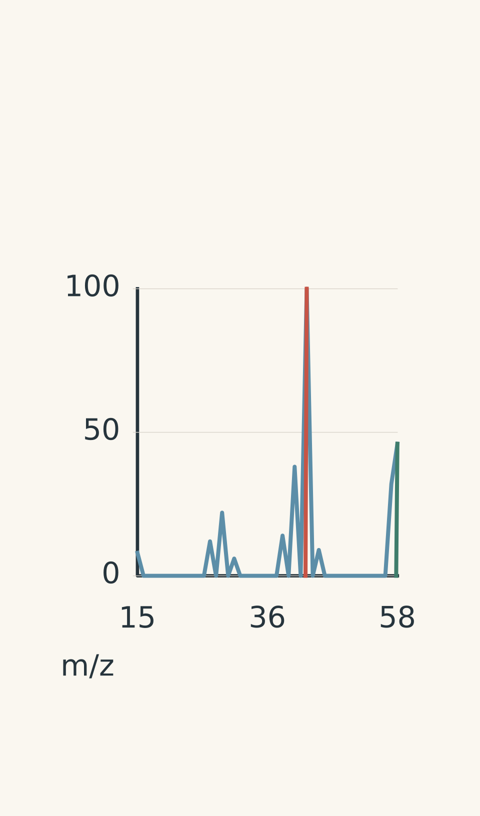
- Representative spectrum
- Base peak — m/z 43
- Molecular ion M⁺• — m/z 58
02The first important peak: the molecular ion
The molecular ion is the original molecule after it loses an electron.
It is often written as \(M^{+\bullet}\). The dot shows that it is a radical ion because it has an unpaired electron.
The molecular ion peak tells us the molecular mass of the compound.
For example, if the molecular ion appears at:
\[ m/z = 58 \]
then the compound has a molecular mass of approximately \(58\ \text{g mol}^{-1}\).
But this is where students often make a mistake.
A common prediction is:
“The biggest peak must be the molecule.”
That feels reasonable because the molecule is the largest piece. However, the biggest peak only tells us which fragment forms most easily. A tiny fragment can be produced in much larger quantities than the original molecular ion.
The molecular ion may be small or even absent if the molecule breaks apart easily.
03Using the molecular ion to find a formula
Once the molecular mass is known, possible molecular formulas can be tested.
For organic compounds containing carbon, hydrogen, oxygen, and nitrogen, the first step is usually checking sensible combinations of atoms.
For example, a molecular ion at \(m/z = 46\) could match:
\[ \ce{C2H6O} \]
because:
\[ (2 \times 12.0) + (6 \times 1.0) + 16.0 = 46.0 \]
The mass fits, but mass spectrometry alone does not always prove one unique structure. Other techniques, such as carbon-13 NMR and proton NMR, provide different structural clues.
You can see how these techniques work together in:
- Carbon-13 NMR Spectroscopy: Interpreting HSC Chemistry Data
- Proton NMR Spectroscopy: How to Interpret HSC Chemistry Data
04Fragment peaks: reading the broken pieces
The real power of mass spectrometry comes from fragments.
Suppose a molecule breaks into:
\[ \ce{CH3+} \]
The fragment has a mass of:
\[ 12.0 + 3(1.0)=15.0 \]
so a peak appears at:
\[ m/z=15 \]
A peak at \(m/z=15\) is often evidence for a methyl group, but it is not proof by itself. Many structures can produce the same small fragments.
The key skill is combining several pieces of evidence.
Think of a detective finding footprints. One footprint is weak evidence. A trail of matching footprints is much stronger.
05A useful analogy: molecular break-ups
A slightly silly way to picture fragmentation is to imagine a group project where everyone gets along differently. If two people always work together, they tend to leave together when the group splits. If a bond in a molecule is easier to break, that part is more likely to leave as a stable fragment.
The analogy helps with the idea of preferred breaking points.
It breaks down because molecules do not choose, have friendships, or make decisions. Fragmentation depends on bond energies, ion stability, and the possible rearrangements of atoms.
06Worked example: finding a molecular formula from a molecular ion
Worked example: A molecular ion at \(m/z=58\)
A mass spectrum of an organic compound shows a molecular ion peak at \(m/z=58\). The compound contains only carbon, hydrogen, and oxygen. Suggest a possible molecular formula.
Step 1
The molecular ion gives:
\[ M = 58\ \text{g mol}^{-1} \]
Step 2
A common starting point is trying carbon atoms because organic compounds usually contain several.
Try two carbon atoms:
\[ 2(12.0)=24.0 \]
Remaining mass:
\[ 58.0-24.0=34.0 \]
Step 3
One oxygen contributes:
\[ 16.0 \]
Remaining:
\[ 34.0-16.0=18.0 \]
which matches six hydrogens:
\[ 6(1.0)=6.0 \]
This leaves too much mass, so try a different combination.
Three carbon atoms:
\[ 3(12.0)=36.0 \]
Remaining:
\[ 58.0-36.0=22.0 \]
One oxygen:
\[ 22.0-16.0=6.0 \]
This gives:
\[ \ce{C3H6O} \]
Step 4
A possible molecular formula is:
\[ \boxed{\ce{C3H6O}} \]
The mass spectrum gives a formula possibility. It does not yet prove the exact arrangement of atoms.
07Worked example: using fragments to distinguish structures
Worked example: A compound has \(M^{+\bullet}=72\) and a strong fragment at \(m/z=43\)
An organic compound has a molecular ion peak at \(m/z=72\). A strong fragment appears at \(m/z=43\). Explain what this suggests.
Step 1
The molecular ion suggests:
\[ M = 72\ \text{g mol}^{-1} \]
A possible formula containing carbon, hydrogen, and oxygen is:
\[ \ce{C4H8O} \]
because:
\[ (4 \times 12.0)+(8 \times 1.0)+16.0=72.0 \]
Step 2
A fragment with mass 43 commonly matches:
\[ \ce{C2H3O+} \]
because:
\[ (2 \times 12.0)+(3 \times 1.0)+16.0=43.0 \]
This fragment is often associated with carbonyl-containing compounds, such as ketones.
Step 3
The molecular ion suggests the overall formula, while the fragment suggests a stable carbonyl-containing fragment.
A possible structure is butanone:
\[ \ce{CH3COCH2CH3} \]
The important reasoning is not memorising “43 means ketone”. The important reasoning is asking:
- What molecular mass fits?
- What fragments are common?
- Which structures could produce those fragments?
08Common mistakes when interpreting mass spectra
| Mistake | Why it seems reasonable | What actually happens |
|---|---|---|
| The tallest peak is the molecular ion | Bigger peak feels like the main molecule | The tallest peak is only the most abundant ion |
| One fragment proves a structure | A fragment looks like a fingerprint | Many compounds can produce the same fragment |
| A molecular mass gives one guaranteed structure | The total number of atoms seems enough | Different arrangements can have the same formula |
| More fragments always means a clearer answer | More data feels better | You need the right fragments, not just more peaks |
Mass spectra are strongest when combined with other structural techniques. Qualitative tests can identify functional groups, while NMR gives information about different chemical environments:
09A practical method for HSC questions
When you are given a mass spectrum, follow this order:
- Find the molecular ion peak if it is present.
- Determine the molecular mass.
- Suggest possible molecular formulas.
- Look at important fragment peaks.
- Match fragments to possible parts of the molecule.
- Check whether the proposed structure explains all major evidence.
Do not start by guessing a structure. Start with constraints.
The best structure is the one that explains the whole spectrum, not just one convenient peak.
10Questions and solutions
Question 1
A mass spectrum of an organic compound shows a molecular ion peak at \(m/z=44\). The compound contains only carbon, hydrogen, and oxygen. Suggest a possible molecular formula.
Solution 1
The possible molecular formula is \(\ce{C2H4O}\).
The molecular ion peak gives:
\[ M=44\ \text{g mol}^{-1} \]
Testing \(\ce{C2H4O}\):
\[ (2 \times 12.0)+(4 \times 1.0)+16.0=44.0 \]
The mass matches, so \(\ce{C2H4O}\) is a possible formula. It is not the only possible structure, because mass spectrometry gives mass information rather than a complete arrangement of atoms.
Question 2
An organic compound has a molecular ion peak at \(m/z=86\). A major fragment appears at \(m/z=43\). Explain why the fragment peak alone cannot identify the compound, and describe what additional reasoning is needed.
Solution 2
The fragment peak at \(m/z=43\) cannot identify the compound by itself because many organic molecules can form fragments with the same mass.
A student might be tempted to say, “The fragment is 43, so the molecule must contain a particular group.” The problem is that different molecules can break in ways that produce identical ions.
The correct approach is to combine evidence:
- the molecular ion gives the total molecular mass, \(86\ \text{g mol}^{-1}\)
- the fragment gives information about a stable piece of the molecule
- the proposed structure must explain both peaks
The fragment is a clue, not a complete answer.
Question 3
A compound has molecular ion \(M^{+\bullet}\) at \(m/z=60\). The spectrum contains strong peaks at \(m/z=45\) and \(m/z=15\). A student claims the compound must be ethanol because the \(m/z=15\) peak represents a methyl group. Explain why this conclusion is not justified.
Solution 3
The conclusion is not justified because a methyl fragment is not unique to ethanol.
The peaks suggest:
- \(m/z=15\) may indicate a \(\ce{CH3+}\) fragment
- \(m/z=45\) may indicate a larger oxygen-containing fragment
However, many compounds can contain a methyl group and produce these fragments.
The molecular ion at \(m/z=60\) must also be considered. A valid structure must have a molecular mass of \(60\ \text{g mol}^{-1}\) and produce the observed fragments.
The trap is treating one fragment as a fingerprint. Mass spectra are interpreted by patterns of evidence.
Question 4
Two compounds have the molecular formula \(\ce{C4H8O}\). Compound A produces a strong \(m/z=43\) peak and compound B does not. Explain why mass spectrometry can distinguish these compounds even though their formulas are identical.
Solution 4
Mass spectrometry can distinguish the compounds because identical formulas do not guarantee identical fragmentation patterns.
Both compounds contain the same atoms, so they have the same molecular mass:
\[ (4 \times 12.0)+(8 \times 1.0)+16.0=72.0\ \text{g mol}^{-1} \]
However, the atoms are arranged differently. When ionised, different bonds are likely to break, producing different stable fragments.
The tempting mistake is thinking that the same formula must create the same spectrum. The missing constraint is molecular structure. The arrangement of atoms controls which fragments form easily.
Question 5
An unknown compound has a molecular ion peak at \(m/z=74\). A student proposes two structures with the same molecular formula:
Structure X: a straight-chain alcohol
Structure Y: an ester with the same number of atoms
The spectrum of the unknown shows a very weak molecular ion peak, a very strong \(m/z=31\) peak, and no obvious \(m/z=43\) peak. Decide which structure is more consistent with the spectrum and explain the reasoning.
Solution 5
Structure X, the straight-chain alcohol, is more consistent with the spectrum.
The molecular ion at \(m/z=74\) suggests both structures could have the same molecular mass, so the molecular ion alone cannot decide.
The key evidence is the fragment pattern:
- a strong \(m/z=31\) peak is consistent with a fragment such as \(\ce{CH2OH+}\), which is commonly associated with alcohol structures
- the weak molecular ion suggests the molecule fragments easily, which is common for some alcohols
- the absence of a strong \(m/z=43\) peak makes a common ester-type fragmentation pathway less supported
The tempting route is to choose either structure because both have the same formula. The deciding constraint is that mass spectrometry measures how the molecule breaks apart, and different arrangements create different preferred fragments.
This is the point where mass spectrometry becomes useful: it does not just tell you what atoms are present. It helps you decide how those atoms are connected. The next step is combining these fragmentation clues with NMR data to build a complete structural picture.